Microscopy – 5 Reasons Coverslips Are Important For High-Quality Imaging

Most people are familiar with coverslips being placed on slides to protect the sample, but that’s not the only reasons that coverslips are important.
They also affect the image quality.
Coverslips function by working with your microscope to focus light to a single point and avoiding unnecessary noise in your image.
Having the wrong type of coverslip will damage the quality of your images and the quality of the data you extract from those images.
So today, we will discuss five reasons why coverslips, and utilizing the right ones, will improve your imaging…
1. Objectives expect there to be a coverslip in the light path.

Objectives are designed to compensate for a coverslip in the light path, and specifically for a 0.17mm thick glass to be in the light path.
This correlates with a number 1.5 coverslip.
You will also find number 1 (0.15mm) and number 2 (0.22mm) coverslips available for sale, but those will not be as effective.
If you check out the Nikon website MicroscopyU, you will see the objective with a numerical aperture of 0.95 will diminish the brightness by almost 80% when there is a variance of only 0.03mm, or a 30 micrometer deviation. Using a #1 coverslip can significantly reduce the brightness of your sample.
Also important to note is that different brands and lines within the brands have different allowable variance within their coverslips, and this becomes important when we’re using high numerical aperture lens or when performing co-localization experiments.
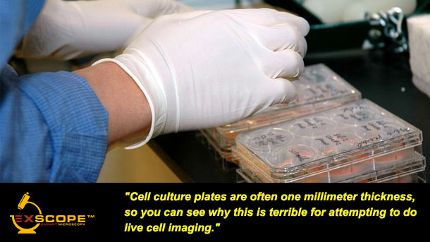
Cell culture plates are often one millimeter thickness, so you can see why this is terrible for attempting to do live cell imaging, or any imaging other than showing the health of your cells.
2. Refractive index is also compensated for within the light path of the objectives.
The refractive index of glass is approximately 1.5, while polystyrene, the most common plastic for tissue culture plates is 1.6.
When we have this mismatch in refractive index, we lose a lot of light through unmatched refraction angles.
This diminishes our signal, so the image becomes more unclear.
You want to get the clearest and most detailed images possible, and to do that you need to ensure that your refractive indexes match up.
This means using plastic tissue culture plates as your imaging coverslip is a bad idea.
3. Thick coverslips take up too much of your working distance.

Many high numerical aperture lenses have a very small working distance where it might only be 300 micrometers or less.
Now, if our coverslip is taking up too much of that, we may not be able to focus through that to our sample.
Similarly, if we have a tissue culture plate that may be one millimeter thick, we physically will not be able to focus through the plate to get to the sample and have an in-focus image.
Too many times, I have seen users put time and resources in preparing their samples, only to find they can’t even focus on their sample.
Why waste your time and money on something as simple as a sample carrier?
Plan ahead and get the right type of coverslip so you can get the images you need the first time.
4. Plastics are depolarizing and light scattering.
This is why we cannot use plastic for DIC imaging, because plastics are depolarizing and scatter light.
If you have apolarizers in a light path you will see rainbows from the stress upon the plastic as well as it will scatter light in many directions, again, losing a loss of signal.
So you end up using more laser power.
But Because you have to up the laser power you might not be able to achieve a good enough signal to noise to have a good image.
5. Plastics introduce autofluorescence.

Many plastics are autofluorescent.
Therefore when we shine a light on the plastic, additional light gets sent back from the sample carrier.
So, if you are trying to get an image of a fluorescent tag or protein in your cells, the autofluorescence of the plastic is going to interfere.
The plastic introduces extra noise into your image and this degrades the quality of the image of your sample.
Which leads to poor results and ultimately a waste of time and money.
It is important to consider the type of coverslip that you are using in your microscopy experiments. Using the wrong kind can have a detrimental effect on the quality of your results. A few of the reasons that the wrong type of coverslip will ruin your experiments are objectives expect there to be a coverslip in the light path, refractive index is also compensated for within the light path of the objectives, thick coverslips take up too much of your working distance, plastics are depolarizing and light scattering, and plastics introduce autofluorescence. Before your next microscopy experiment, double check that you are using the right kind of coverslip.
To learn more about Microscopy – 5 Reasons Coverslips Are Important For High-Quality Imaging, and to get access to all of our advanced microscopy materials including training videos, presentations, workbooks, and private group membership, get on the Expert Microscopy wait list.

ABOUT HEATHER BROWN-HARDING
Heather Brown-Harding, PhD, is currently the assistant director of Wake Forest Microscopy and graduate teaching faculty.She also maintains a small research group that works on imaging of host-pathogen interactions. Heather is passionate about making science accessible to everyone.High-quality research shouldn’t be exclusive to elite institutions or made incomprehensible by unnecessary jargon. She created the modules for Excite Microscopy with this mission.
In her free time, she enjoys playing with her cat & dog, trying out new craft ciders and painting.You can find her on twitter (@microscopyEd) a few times of day discussing new imaging techniques with peers.
More Written by Heather Brown-Harding


