3 Experiments You Can Do Easier On An ImageStream Flow Cytometer

What happens if one combines the power and speed of traditional flow cytometers with the resolution of a microscope?

Cytometry is the study of biological processes at the whole cell level and includes techniques like light microscopy and electron microscopy.

But microscopy by itself is a bit different.

From the earliest days of microscopy, including the use of the first true microscopes by van Leeuwenhoek and others, scientists have been able to start seeing the finest details of a cell.

With the development of the flow cytometer, researchers have been able to explore cellular processes in great deal.  For example, modern flow cytometry technology allows for the measure 14 or more parameters at the same time.

However, one of the limitations of traditional flow cytometry is that these fine details of the cell are lost.

Bringing Flow Cytometry And Microscopy Together

Combining the power and speed of traditional flow cytometers with the resolution of a microscope has been the goal of many for over 20 years.

The first flow cytometry-microscopy hybrid system was described in the late 1970’s in a paper by Leon Wheeless.

The Wheeless system looked something like this:

Wheeless System Flow Cytometry Miscroscopy

It took over 20 years for this technology to be expanded and commercialized into an instrument called the ImageStream.

The below figure summarizes the power of the ImageStream and how it brings together flow cytometry and traditional light microscopy (morphology).

Image Stream

ImageStream technology allows for complex morphometric measurements in a phenotypically defined way at rates of 100 cells per second.

ImageStream technology simplifies many experiments and often provides for more robust results of these experiments.

Here are 3 such experiments made easier and better by ImageStream technology:

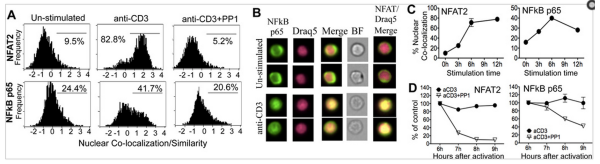
1. Measuring Nuclear Translocation Events

It is well known that the translocation of proteins like NF-kB from the cytoplasm to the nucleus have dramatic results on the cell through changing expression of target genes.

Using the Imagestream, it is possible to co-localize two fluorescent signals using the Similarity Index that researchers in the Fowell lab developed, allowing them to dissect the regulatory mechanisms of NFAT and NF-kB signaling resulting in Treg cells attenuating IL-2 production by CD4 cells.

As the below figure demonstrates, it is possible to identify cells that are co-localizing proteins in the nucleus using the ImageStream.

ExCyte Co-Localization ImageStream

How robust is this method?

Minderman and coworkers did a correlative study using Imagestream, traditional microscopy, and Western blot analysis and concluded:

It is demonstrated that ImageStream assessment of receptor-mediated (TNFα) and drug (Daunorubicin, DNR)-induced NF-κB translocation in leukemic cell lines correlates well with microscopy analysis and Western blot analysis. It is further demonstrated that ImageStream cytometry enables quantitative assessment of p65 translocation in immunophenotypically-defined subpopulations; and that this assessment is highly reproducible.

2. Characterizing Cellular Morphology

Cell biology literature is full of morphological characterization of cells.

Flow cytometry takes that characterization to a different level, using expressed proteins as a means of identifying the cells of interest.

With the ImageStream, it is possible to combine these morphological measures with fluorescent measures to get a better understanding of the overall process.

This is elegantly demonstrated in a paper by Kathleen McGrath and Jim Palis in the area of red blood cell development .

Using three fluorescent probes and morphological characteristics from the rich literature on red blood cells, they were able to develop a gating strategy to characterize the development of the RBC from pro-erythroblast to mature red blood cells, (Figure 3 from their paper is shown below).

Image Stream

With this foundation, McGrath and Palis’ groups have been able to extend the use of the ImageStream to identify the pyrenocyte (or nucleated cell with a rim of cytoplasm left over from RBC enucleation events), understand the effects of sub-lethal radiation on the RBC development and, expand the characterization of megakaryocytes.

Combining morphology and fluorescence cytometry really opened up new avenues of investigation for these researchers and it can for you too.

3. Rare Event Analysis

One limitation of traditional flow cytometry is that very often cells will bind to parts of other cells, forming doublets.

The problem is that there is not enough resolution to ensure that the cell that’s marked as a doublet is in fact a doublet and not a larger cell.

This issue can have profound effects when trying to measure rare events.

Take the case of circulating endothelial cells, which have an estimated frequency of 1 to 10 per ml of blood. In a paper by Samsel (in the lab of Phil McCoy), the researchers successfully used a rare CEC to measure the efficiency of detection of these cells with the ImageStream.

The ImageStream also allowed them to successfully inspect of the cells that were being excluded.

As shown in Figure 4 of the researcher’s paper, the excluded cells were visualized.

Image Stream Rare Cells

These are three very different areas of research that are impacted by having the ability to see the fine details of the cells that are being characterized.

The ImageStream opens up new areas of investigation and allows for new complex questions to be answered.

To learn more about automatic compensation and to get access to all of our advanced materials including 20 training videos, presentations, workbooks, and private group membership, get on Mastery Class wait list.

Join Expert Cytometry's Mastery Class

ABOUT TIM BUSHNELL, PHD

Tim Bushnell holds a PhD in Biology from the Rensselaer Polytechnic Institute. He is a co-founder of—and didactic mind behind—ExCyte, the world’s leading flow cytometry training company, which organization boasts a veritable library of in-the-lab resources on sequencing, microscopy, and related topics in the life sciences.

Tim Bushnell, PhD

Similar Articles

The Power Of Spectral Viewers And Their Use In Full Spectrum Flow Cytometry

The Power Of Spectral Viewers And Their Use In Full Spectrum Flow Cytometry

By: Tim Bushnell, PhD

What photon from yonder fluorochrome breaks?  It is … umm… hmmm. Let me see. Excitation off a 561 nm laser, with an emission maximum of 692 nm. I’m sure if Shakespeare was a flow cytometrist, he might have written that very scene. But the play is lost in time. However, since the protagonist had difficulty determining what fluorochrome was emitting photons, let’s consider how this could be figured out. In my opinion, one of the handiest flow cytometry tools is the spectral viewer. This tool helps visualize the excitation and emission profile of different fluorochromes, as well as allowing you…

3 Must-Have High-Dimensional Flow Cytometry Controls

3 Must-Have High-Dimensional Flow Cytometry Controls

By: Tim Bushnell, PhD

Developments such as the recent upgrade to the Cytobank analysis platform and the creation of new packages such as Immunocluster are reducing the computational expertise needed to work with high-dimensional flow cytometry datasets. Whether you are a researcher in academia, industry, or government, you may want to take advantage of the reduced barrier to entry to apply high-dimensional flow cytometry in your work. However, you’ll need the right experimental design to access the new transformative insights available through these approaches and avoid wasting the considerable time and money required for performing them. As with all experiments, a good design begins…

The Fluorochrome Less Excited: How To Build A Flow Cytometry Antibody Panel

The Fluorochrome Less Excited: How To Build A Flow Cytometry Antibody Panel

By: Tim Bushnell, PhD

Fluorochrome, antibodies and detectors are important. The journey of a thousand cells starts with a good fluorescent panel. The polychromatic panel is the combination of antibodies and fluorochromes. These will be used during the experiment to answer the biological question of interest. When you only need a few targets, the creation of the panel is relatively straightforward. It’s only when you start to get into more complex panels with multiple fluorochromes that overlap in excitation and emission gets more interesting.  FLUOROCHROMES Both full spectrum and traditional fluorescent flow cytometry rely on measuring the emission of the fluorochromes that are attached…

Flow Cytometry Year in Review: Key Changes To Know

Flow Cytometry Year in Review: Key Changes To Know

By: Meerambika Mishra

Here we are, at the end of an eventful year 2021. But with the promise of a new year 2022 to come. It has been a long year, filled with ups and downs. It is always good to reflect on the past year as we move to the future.  In Memoriam Sir Isaac Newton wrote “If I have seen further, it is by standing upon the shoulders of giants.” In the past year, we have lost some giants of our field including Zbigniew Darzynkiwicz, who contributed much in the areas of cell cycle analysis and apoptosis. Howard Shapiro, known for…

What Star Trek Taught Me About Flow Cytometry

What Star Trek Taught Me About Flow Cytometry

By: Tim Bushnell, PhD

It is no secret that I am a very big fan of the Star Trek franchise. There are many good episodes and lessons explored in the 813+ episodes, 12 movies (and counting). Don’t worry, this blog is not going to review all 813, or even 5 of them. Instead, some of the lessons I have taken away from the show that have applicability to science and flow cytometry.  “Darmok and Jalad at Tanagra.”  (ST:TNG season 5, episode 2) This is probably one of my favorite episodes, which involves Picard and an alien trying to establish a common ground and learn…

5 Flow Cytometry Strategies That Sun Tzu Taught Me

5 Flow Cytometry Strategies That Sun Tzu Taught Me

By: Tim Bushnell, PhD

Sun Tzu was a Chinese general and philosopher. His most famous writing is ‘The Art of War’, and has been studied by generals and CEOs, to glean ideas and strategies to help their missions. I was recently rereading this work and thought to myself if any of Sun Tzu’s lessons could apply to flow cytometry.  So I have identified 5 points that I think lend themselves to thinking about flow cytometry.  “Quickness is the essence of the war.” In flow cytometry, speed is of the essence. The longer the cells are out of their natural environment, the less happy they…

A Basic Guide To Flow Cytometry (3 Foundational Concepts)

A Basic Guide To Flow Cytometry (3 Foundational Concepts)

By: Meerambika Mishra

Mastering foundational concepts are imperative for successfully using any technique or system.  Robert Heinlein introduced the term ‘Grok’  in his novel Stranger in a Strange Land. Ever since then it has made its way into popular culture. To Grok something is to understand it intuitively, fully. As a cytometrist, there are several key concepts that you must grok to be successful in your career. These foundational concepts are the key tools that we use day in and day out to identify and characterize our cells of interest.  Cells Flow cytometry measures biological processes at the whole cell level. To do…

Which Fluorophores To Use For Your Microscopy Experiment

Which Fluorophores To Use For Your Microscopy Experiment

By: Heather Brown-Harding, PhD

Fluorophore selection is important. I have often been asked by my facility users which fluorophore is best suited for their experiments. The answer to this is mostly dependent on whether they are using a widefield microscope with set excitation/emission cubes or a laser based system that lets you select the laser and the emission window. Once you have narrowed down which fluorophores you can excite and collect the correct emission, you can further refine the specific fluorophore that is best for your experiment.  In this blog  we will discuss how to determine what can work with your microscope, and how…

4 No Cost Ways To Improve Your Microscopy Image Quality

4 No Cost Ways To Improve Your Microscopy Image Quality

By: Heather Brown-Harding, PhD

Image quality is critical for accurate and reproducible data. Many people get stuck on the magnification of the objective or on using a confocal instead of a widefield microscope. There are several other factors that affect the image quality such as the numerical aperture of the objective, the signal-to-noise ratio of the system, or the brightness of the sample.  Numerical aperture is the ability of an objective to collect light from a sample, but it contributes to two key formulas that will affect your image quality. The first is the theoretical resolution of the objective. It is expressed with the…

Top Industry Career eBooks

Get the Advanced Microscopy eBook

Get the Advanced Microscopy eBook

Heather Brown-Harding, PhD

Learn the best practices and advanced techniques across the diverse fields of microscopy, including instrumentation, experimental setup, image analysis, figure preparation, and more.

Get The Free Modern Flow Cytometry eBook

Get The Free Modern Flow Cytometry eBook

Tim Bushnell, PhD

Learn the best practices of flow cytometry experimentation, data analysis, figure preparation, antibody panel design, instrumentation and more.

Get The Free 4-10 Compensation eBook

Get The Free 4-10 Compensation eBook

Tim Bushnell, PhD

Advanced 4-10 Color Compensation, Learn strategies for designing advanced antibody compensation panels and how to use your compensation matrix to analyze your experimental data.