Why You Need To Use FMO Controls For All Multicolor Flow Cytometry Experiments

What are the three most important parts of any flow cytometry experiment?

Controls, controls, controls.

Of course, this is mostly in jest. Mostly.

Reagent selection, well-maintained instruments, and other factors are also important. However, you must control for these factors.

Setting up the right controls is critical to determining how your cells are responding to treatment. It’s also important for correctly interpreting your data and drawing correct conclusions. Without the proper controls, you would not be able to compensate your flow cytometry experiments correctly or identify your cells of interest correctly.

By using the wrong controls, or leaving out certain controls altogether, you leave yourself wide open for criticism. Your gate placement will be questioned, your samples will be questioned, and your papers and grants will be questioned.

Why Fluorescence Minus One, Or FMO Controls, Are Important

We’ve all had experience working with someone who wanted to see the “real” samples, not the control samples, first.

But if you’re a smart scientist, you know “real,” or experimental samples are meaningless unless you know, among other things, the background levels you’re working with.

Valuing your control samples over your experimental samples is a mistake, especially when performing flow cytometry experiments. For example, in flow cytometry, some gates are “easy” to set. CD19+ B cells are often easy to pick out on a plot showing CD3 for a T cell subset sample. Other gates, however, are more difficult to define. This is especially true when you’re looking at activation markers within a continuum, or dim levels of positivity, like FoxP3 in regulatory T cells. How can you convince reviewers that you didn’t make an error and placed your gate in the proper place?

Now, consider gating out subsets in complicated 10+ color experiments. How are you going to account for the data spread that occurs with compensation? In any multicolor flow cytometry experiment, the answer to your gating troubles is to use Fluorescence Minus One, or FMO, controls.

What Is A Fluorescence Minus One, Or FMO, Control?

FMO controls are samples that contain all the antibodies you are testing in your experimental samples, minus one of them.

When analyzing the minus, or left out parameter in an FMO control, you give yourself a strong negative control to work with. It’s a strong negative control because the left out marker in the FMO control allows you to take into account how the other stains in your panel affect the left out parameter.

FMO controls are required for accurately discriminating positive versus negative signals, high versus low (or variable) antigen expression levels, and more. Even simple 2- or 3-color experiments reveal the need for FMO controls when drawing gates.

The figure below shows data from an experiment that was set up to identify naive versus memory CD4+ T cells. After properly compensating the data, the experimenter is left with the plot on the far right. Now the question becomes, where does the experimenter set the gate for PE positivity?

If the experimenter uses an unstained control only (far left panel), he or she is able to set a boundary (red dashed line) for PE positivity. However, if you now look at the middle plot, you can see that the bulk of the cells are labeled as “positive” for PE because they are above the boundary established by the unstained control.

multicolor flow cytometry training | Expert Cytometry | fmo control flow cytometry

Notice that the cells in the middle plot are stained with all the fluorochromes in the experimental samples except PE. As a result, they can NOT be positive for PE. This middle panel represents the FMO control in this experiment and shows the experimenter that the true negative boundary for PE, or FMO bound, is where the blue line lies.

Now, using the FMO control and resulting FMO bound, the experimenter is able to set the proper gate and ensure that the spread of the data (blue double arrow on the far right), is not impacting his or her gating.

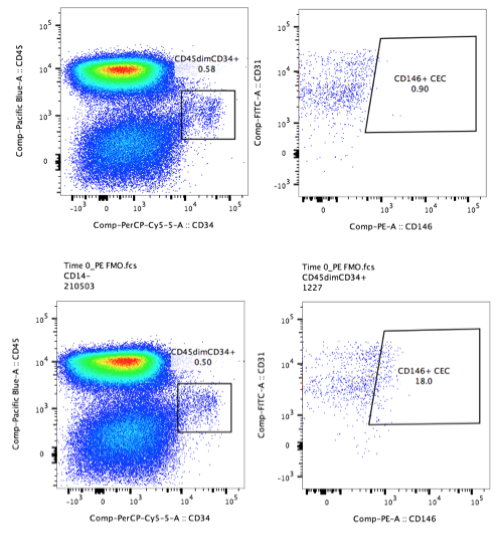
In the more complex example below, the plots show a population of circulating CD34+ cells in human PBMCs. The experimenter who ran these samples wanted to determine the levels of CD146 on these cells. The experimenter included FMO controls when testing the panel (CD146 on PE and CD31 on FITC, bottom two plots). As a result, the experimenter was easily able to draw gates around the populations of interest despite low levels of background positivity (top two plots).

multicolor flow cytometry training | Expert Cytometry | fmo control flow cytometry

When To Use Fluorescence Minus One, Or FMO Controls

  1. When running any multicolor flow cytometry experiment, there is a spread in the data that quickly becomes apparent when you compensate your data. Due to the physics of fluorescence and the exponential scales used to display flow cytometry data, this spread is unavoidable. However, using FMO controls in your flow cytometry experiments can remove all ambiguity from your compensated multicolor plots.
  2. When antigens are dim or variably expressed, the use of FMO controls is critical. This is especially true when analyzing cell activation markers on immune cells. (Don’t forget those unstimulated biological controls here either!)
  3. Do NOT add isotype antibodies to your FMO controls. These antibodies do NOT add any useful information to the FMO. If you are working with myeloid cells and think you need an isotype control, prepare it separately.
  4. If you are unsure of the levels of expression you are dealing with in your samples or the sensitivity of your antibodies or samples, use FMO controls. It’s better to prepare a few controls you may not need than lose money, time, publications or grant funding by being stingy.

When in doubt, follow Maecker’s advice reported in Cytometry, “When high-quality monoclonal antibody conjugates are used at appropriate concentrations, they tend to have relatively low background staining. As such, in experiments of >4 colors, the major source of background staining tends to be fluorescence spillover. Because of this, the use of FMO controls has become both popular and prudent.”

Fluorescence Minus One, or FMO controls are critical for any scientist who wants to back up his or her drawing of flow cytometry gates. When you use FMO controls, you establish scientific evidence as to why the gates you drew are drawn correctly. This is true for any multicolor flow cytometry experiment. FMO controls will give you, as well as those who view your work with a critical eye, confidence in the degree of accuracy of your measurements.

To learn more about FMO controls, and to get access to all of our advanced materials including 20 training videos, presentations, workbooks, and private group membership, get on the Flow Cytometry Mastery Class wait list.

Join Expert Cytometry's Mastery Class

ABOUT TIM BUSHNELL, PHD

Tim Bushnell holds a PhD in Biology from the Rensselaer Polytechnic Institute. He is a co-founder of—and didactic mind behind—ExCyte, the world’s leading flow cytometry training company, which organization boasts a veritable library of in-the-lab resources on sequencing, microscopy, and related topics in the life sciences.

Tim Bushnell, PhD

Similar Articles

Fickle Markers: Solutions For Antibody Binding Specificity Challenges

Fickle Markers: Solutions For Antibody Binding Specificity Challenges

By: Tim Bushnell, PhD

Reproducibility has been an ongoing, and important, concept in the sciences for years.  In the area of biomedical research, the alarm was sounded by several papers published in the early 2010’s.  Authors like Begley and Ellis, Prinz and coworkers, and Vasilevsky and colleagues, among others reported an alarming trend in the reproducibility of pre-clinical data.  These reports indicated between 50% to almost 90% of published pre-clinical data were not reproducible.  This was further highlighted in the article by Freedman and coworkers, who tried to identify and quantify the different sources of error that could be causing this crisis.  Figure 1,…

The Fluorochrome Less Excited: How To Build A Flow Cytometry Antibody Panel

The Fluorochrome Less Excited: How To Build A Flow Cytometry Antibody Panel

By: Tim Bushnell, PhD

Fluorochrome, antibodies and detectors are important. The journey of a thousand cells starts with a good fluorescent panel. The polychromatic panel is the combination of antibodies and fluorochromes. These will be used during the experiment to answer the biological question of interest. When you only need a few targets, the creation of the panel is relatively straightforward. It’s only when you start to get into more complex panels with multiple fluorochromes that overlap in excitation and emission gets more interesting.  FLUOROCHROMES Both full spectrum and traditional fluorescent flow cytometry rely on measuring the emission of the fluorochromes that are attached…

Flow Cytometry Year in Review: Key Changes To Know

Flow Cytometry Year in Review: Key Changes To Know

By: Meerambika Mishra

Here we are, at the end of an eventful year 2021. But with the promise of a new year 2022 to come. It has been a long year, filled with ups and downs. It is always good to reflect on the past year as we move to the future.  In Memoriam Sir Isaac Newton wrote “If I have seen further, it is by standing upon the shoulders of giants.” In the past year, we have lost some giants of our field including Zbigniew Darzynkiwicz, who contributed much in the areas of cell cycle analysis and apoptosis. Howard Shapiro, known for…

What Star Trek Taught Me About Flow Cytometry

What Star Trek Taught Me About Flow Cytometry

By: Tim Bushnell, PhD

It is no secret that I am a very big fan of the Star Trek franchise. There are many good episodes and lessons explored in the 813+ episodes, 12 movies (and counting). Don’t worry, this blog is not going to review all 813, or even 5 of them. Instead, some of the lessons I have taken away from the show that have applicability to science and flow cytometry.  “Darmok and Jalad at Tanagra.”  (ST:TNG season 5, episode 2) This is probably one of my favorite episodes, which involves Picard and an alien trying to establish a common ground and learn…

5 Flow Cytometry Strategies That Sun Tzu Taught Me

5 Flow Cytometry Strategies That Sun Tzu Taught Me

By: Tim Bushnell, PhD

Sun Tzu was a Chinese general and philosopher. His most famous writing is ‘The Art of War’, and has been studied by generals and CEOs, to glean ideas and strategies to help their missions. I was recently rereading this work and thought to myself if any of Sun Tzu’s lessons could apply to flow cytometry.  So I have identified 5 points that I think lend themselves to thinking about flow cytometry.  “Quickness is the essence of the war.” In flow cytometry, speed is of the essence. The longer the cells are out of their natural environment, the less happy they…

A Basic Guide To Flow Cytometry (3 Foundational Concepts)

A Basic Guide To Flow Cytometry (3 Foundational Concepts)

By: Meerambika Mishra

Mastering foundational concepts are imperative for successfully using any technique or system.  Robert Heinlein introduced the term ‘Grok’  in his novel Stranger in a Strange Land. Ever since then it has made its way into popular culture. To Grok something is to understand it intuitively, fully. As a cytometrist, there are several key concepts that you must grok to be successful in your career. These foundational concepts are the key tools that we use day in and day out to identify and characterize our cells of interest.  Cells Flow cytometry measures biological processes at the whole cell level. To do…

Which Fluorophores To Use For Your Microscopy Experiment

Which Fluorophores To Use For Your Microscopy Experiment

By: Heather Brown-Harding, PhD

Fluorophore selection is important. I have often been asked by my facility users which fluorophore is best suited for their experiments. The answer to this is mostly dependent on whether they are using a widefield microscope with set excitation/emission cubes or a laser based system that lets you select the laser and the emission window. Once you have narrowed down which fluorophores you can excite and collect the correct emission, you can further refine the specific fluorophore that is best for your experiment.  In this blog  we will discuss how to determine what can work with your microscope, and how…

4 No Cost Ways To Improve Your Microscopy Image Quality

4 No Cost Ways To Improve Your Microscopy Image Quality

By: Heather Brown-Harding, PhD

Image quality is critical for accurate and reproducible data. Many people get stuck on the magnification of the objective or on using a confocal instead of a widefield microscope. There are several other factors that affect the image quality such as the numerical aperture of the objective, the signal-to-noise ratio of the system, or the brightness of the sample.  Numerical aperture is the ability of an objective to collect light from a sample, but it contributes to two key formulas that will affect your image quality. The first is the theoretical resolution of the objective. It is expressed with the…

What Is Total Internal Reflection Fluorescence (TIRF) Microscopy & Is It Right For You?

What Is Total Internal Reflection Fluorescence (TIRF) Microscopy & Is It Right For You?

By: Heather Brown-Harding, PhD

TIRF is not as common as other microscopy based techniques due to certain restrictions. We will discuss these restrictions, then analyze why it might be perfect for your experiment.  TIRF relies on an evanescent wave, created through a critical angle of coherent light (i.e. laser) that reaches a refractive index mismatch.  What does it mean in practice?  A high angle laser reflects off the interface of the coverslip and the sample. Although the depth that this wave penetrates is dependent on the wavelength of the light, in practice it is approximately 50-300nm from the coverslip. Therefore, the cell membrane is…

Top Industry Career eBooks

Get the Advanced Microscopy eBook

Get the Advanced Microscopy eBook

Heather Brown-Harding, PhD

Learn the best practices and advanced techniques across the diverse fields of microscopy, including instrumentation, experimental setup, image analysis, figure preparation, and more.

Get The Free Modern Flow Cytometry eBook

Get The Free Modern Flow Cytometry eBook

Tim Bushnell, PhD

Learn the best practices of flow cytometry experimentation, data analysis, figure preparation, antibody panel design, instrumentation and more.

Get The Free 4-10 Compensation eBook

Get The Free 4-10 Compensation eBook

Tim Bushnell, PhD

Advanced 4-10 Color Compensation, Learn strategies for designing advanced antibody compensation panels and how to use your compensation matrix to analyze your experimental data.