When To Use (And Not Use) Flow Cytometry Isotype Controls

Antibodies can bind to cells in a specific manner – where the FAB portion of the antibody binds to a high-affinity specific target or the FC portion of the antibody binds to the FcR on the surface of some cells.

They can also bind to cells in a nonspecific manner, where the FAB portion binds to a low affinity, non-specific target. Further, as cells die, and the membrane integrity is compromised, antibodies can non-specifically bind to intracellular targets.

The question has always been how to identify and control for the nonspecific antibody binding observed.  

This led to many research groups using a control called the Isotype control.

The concept of this control is that an antibody targeting a protein not on the surface of the target cells, has the same isotype (both heavy and light chain) as the antibody of interest.  When used to label cells, those that showed binding to the isotype, would be excluded as they represented the non-specific binding of the cells.

When Isotype Controls Were Everything

Isotype controls were once THE negative control for flow cytometry experiments.

They are still very often included by some labs, almost abandoned by others, and a subject of confusion for many beginners. What are they, why and when do I need them? Are they of any use at all, or just a waste of money?

Most importantly, why do reviewers keep asking for them when they review papers containing flow data?

Isotype controls were classically meant to show what level of nonspecific binding you might have in your experiment. The idea is that there are several ways that an antibody might react in undesirable ways with the surface of the cell.

Not all of these can be directly addressed by this control (such as cross-reactivity to a similar epitope on a different antigen, or even to a different epitope on the same antigen). What it does do is give you an estimate of non-specific (non-epitope-driven) binding. This can be Fc mediated binding, or completely nonspecific “sticky” cell adhesion.

In order to be useful, the isotype control should ideally be the same isotype, both in terms of species, heavy chain (IgA, IgG, IgD, IgE, or IgM) and light chain (kappa or lambda) class, the same fluorochrome (PE, APC, etc.), and have the same F:P ratio. F:P is a measurement of how many fluorescent molecules are present on each antibody.

This, unfortunately, makes the manufacture of ideal isotype controls highly impractical. 

There is even a case to be made that differences in the amino acid sequence of the variable regions of both the light and heavy chains might result in variable levels of undesirable adherence in isotypes versus your antibody of interest.

Moving Beyond Isotype Controls

For these reasons, many in the field are moving beyond the isotype control. With some suggesting they be left out of almost all experiments.

If you spend any time browsing the Purdue Cytometry list, you’ll see these same arguments presented in threads about isotype controls. 

The following paper presents options for controls in several categories, the options available, and pros and cons of each option. The section on isotype controls summarizes the problems with the use of isotype controls very clearly…

Flow cytometry controls, instrument setup, and the determination of positivity.

Additionally, the following paper presents options for controls in several categories, the options available, and pros and cons of each option…

Considerations for the control of background fluorescence in clinical flow cytometry.

The section of the above paper focusing on isotype controls summarizes the problems with their use very clearly.

The article also illustrates difference in undesirable binding at different levels using the same clone from different manufacturers.

See the figure below for an example of how even the same isotype control clone can result in highly variable levels of undesirable staining.

Isotype Antibody | Expert Cytometry | Fluoresence

If you do use isotype controls in your experiment, they must match as many of the following characteristics as possible for your specific antibody—species, isotype, fluorochrome, F:P ratio, and concentration.

9 Tips For Using (Or Not Using) Isotype Controls

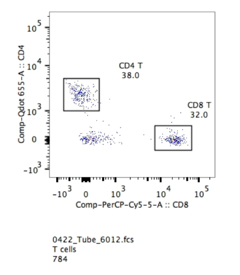
1. You certainly don’t need them for things that are clearly bimodal. If you are looking for T cells and B cells in peripheral blood the negative cells also in the circulation provide gating confidence. As seen in the example below, it is extremely easy to pick out CD4 and CD8 positive cells in the sample of lysed mouse blood.

Isotype Matched Control | Expert Cytometry | igg Isotype


2. If you are using post-cultured cells, the isotype control might give you some information about the inherent “stickiness” of your cells. However, this is not meant as a value you can subtract from your specific antibody sample in terms of fluorescence intensity or percent positive. This is simply a qualitative measure of “stickiness” and the effectiveness of blocking in your protocol.

3. If you are using multiple dyes in your search, and your concern is positivity by spectral overlap, you will be better served by using a fluorescence-minus-one control (FMO), in which all antibodies are included except the one you suspect is most prone to error from spectral overlap.

4. You should absolutely not be using them to determine positive versus negative cells, or as a gating control in your assay.

5. Keep in mind that the best way to avoid high levels of background staining of antigen-negative populations is to carefully titrate your reagents to ensure the highest positive signals in bright populations, while reducing spread in the negative population.

6. If you are using your isotype control and you are seeing high levels of non-desired staining, it is time to look carefully at your blocking step in your protocol. Are you using an Fc-block if you have myeloid cells? Have you tried adding excess immunoglobulins or whole serum to your buffer? Each of these can help pull down your nonspecific adherence.

7. Are you certain it is non-specific antibody adherence you are are dealing with and not free fluorochrome adherence? You can find out by using an isoclonic control. If you add massive amounts of non-fluorochrome conjugated monoclonal antibody to your staining reaction, your fluorescence should drop. If it does not, your issue is not due to nonspecific antibody binding, but to free fluorochrome binding.

8. For cell signalling and cytokine staining, besides the FMO control, make sure not to neglect a biological negative control, whether that be unstimulated cells, or cells treated with an inhibitor of phosphorylation.

9. Don’t forget to use a viability dye in the polychromatic panel. With the proliferation of these dyes in different colors and for both viable and fixed cells, there is no reason to not use these dyes. Viability dyes are essential for removing dead cells that will non-specifically uptake antibodies.

In conclusion, isotype controls are useful for demonstrating that there was poor blocking of the cells. They should never be used to determine or set positivity in fully stained samples.

To learn more about which controls you should use for your flow cytometry experiments and to get access to all of our advanced materials including 20 training videos, presentations, workbooks, and private group membership, get on the Flow Cytometry Mastery Class wait list.

Join Expert Cytometry's Mastery Class

ABOUT TIM BUSHNELL, PHD

Tim Bushnell holds a PhD in Biology from the Rensselaer Polytechnic Institute. He is a co-founder of—and didactic mind behind—ExCyte, the world’s leading flow cytometry training company, which organization boasts a veritable library of in-the-lab resources on sequencing, microscopy, and related topics in the life sciences.

Tim Bushnell, PhD

Similar Articles

Fickle Markers: Solutions For Antibody Binding Specificity Challenges

Fickle Markers: Solutions For Antibody Binding Specificity Challenges

By: Tim Bushnell, PhD

Reproducibility has been an ongoing, and important, concept in the sciences for years.  In the area of biomedical research, the alarm was sounded by several papers published in the early 2010’s.  Authors like Begley and Ellis, Prinz and coworkers, and Vasilevsky and colleagues, among others reported an alarming trend in the reproducibility of pre-clinical data.  These reports indicated between 50% to almost 90% of published pre-clinical data were not reproducible.  This was further highlighted in the article by Freedman and coworkers, who tried to identify and quantify the different sources of error that could be causing this crisis.  Figure 1,…

The Fluorochrome Less Excited: How To Build A Flow Cytometry Antibody Panel

The Fluorochrome Less Excited: How To Build A Flow Cytometry Antibody Panel

By: Tim Bushnell, PhD

Fluorochrome, antibodies and detectors are important. The journey of a thousand cells starts with a good fluorescent panel. The polychromatic panel is the combination of antibodies and fluorochromes. These will be used during the experiment to answer the biological question of interest. When you only need a few targets, the creation of the panel is relatively straightforward. It’s only when you start to get into more complex panels with multiple fluorochromes that overlap in excitation and emission gets more interesting.  FLUOROCHROMES Both full spectrum and traditional fluorescent flow cytometry rely on measuring the emission of the fluorochromes that are attached…

Flow Cytometry Year in Review: Key Changes To Know

Flow Cytometry Year in Review: Key Changes To Know

By: Meerambika Mishra

Here we are, at the end of an eventful year 2021. But with the promise of a new year 2022 to come. It has been a long year, filled with ups and downs. It is always good to reflect on the past year as we move to the future.  In Memoriam Sir Isaac Newton wrote “If I have seen further, it is by standing upon the shoulders of giants.” In the past year, we have lost some giants of our field including Zbigniew Darzynkiwicz, who contributed much in the areas of cell cycle analysis and apoptosis. Howard Shapiro, known for…

What Star Trek Taught Me About Flow Cytometry

What Star Trek Taught Me About Flow Cytometry

By: Tim Bushnell, PhD

It is no secret that I am a very big fan of the Star Trek franchise. There are many good episodes and lessons explored in the 813+ episodes, 12 movies (and counting). Don’t worry, this blog is not going to review all 813, or even 5 of them. Instead, some of the lessons I have taken away from the show that have applicability to science and flow cytometry.  “Darmok and Jalad at Tanagra.”  (ST:TNG season 5, episode 2) This is probably one of my favorite episodes, which involves Picard and an alien trying to establish a common ground and learn…

5 Flow Cytometry Strategies That Sun Tzu Taught Me

5 Flow Cytometry Strategies That Sun Tzu Taught Me

By: Tim Bushnell, PhD

Sun Tzu was a Chinese general and philosopher. His most famous writing is ‘The Art of War’, and has been studied by generals and CEOs, to glean ideas and strategies to help their missions. I was recently rereading this work and thought to myself if any of Sun Tzu’s lessons could apply to flow cytometry.  So I have identified 5 points that I think lend themselves to thinking about flow cytometry.  “Quickness is the essence of the war.” In flow cytometry, speed is of the essence. The longer the cells are out of their natural environment, the less happy they…

A Basic Guide To Flow Cytometry (3 Foundational Concepts)

A Basic Guide To Flow Cytometry (3 Foundational Concepts)

By: Meerambika Mishra

Mastering foundational concepts are imperative for successfully using any technique or system.  Robert Heinlein introduced the term ‘Grok’  in his novel Stranger in a Strange Land. Ever since then it has made its way into popular culture. To Grok something is to understand it intuitively, fully. As a cytometrist, there are several key concepts that you must grok to be successful in your career. These foundational concepts are the key tools that we use day in and day out to identify and characterize our cells of interest.  Cells Flow cytometry measures biological processes at the whole cell level. To do…

Which Fluorophores To Use For Your Microscopy Experiment

Which Fluorophores To Use For Your Microscopy Experiment

By: Heather Brown-Harding, PhD

Fluorophore selection is important. I have often been asked by my facility users which fluorophore is best suited for their experiments. The answer to this is mostly dependent on whether they are using a widefield microscope with set excitation/emission cubes or a laser based system that lets you select the laser and the emission window. Once you have narrowed down which fluorophores you can excite and collect the correct emission, you can further refine the specific fluorophore that is best for your experiment.  In this blog  we will discuss how to determine what can work with your microscope, and how…

4 No Cost Ways To Improve Your Microscopy Image Quality

4 No Cost Ways To Improve Your Microscopy Image Quality

By: Heather Brown-Harding, PhD

Image quality is critical for accurate and reproducible data. Many people get stuck on the magnification of the objective or on using a confocal instead of a widefield microscope. There are several other factors that affect the image quality such as the numerical aperture of the objective, the signal-to-noise ratio of the system, or the brightness of the sample.  Numerical aperture is the ability of an objective to collect light from a sample, but it contributes to two key formulas that will affect your image quality. The first is the theoretical resolution of the objective. It is expressed with the…

What Is Total Internal Reflection Fluorescence (TIRF) Microscopy & Is It Right For You?

What Is Total Internal Reflection Fluorescence (TIRF) Microscopy & Is It Right For You?

By: Heather Brown-Harding, PhD

TIRF is not as common as other microscopy based techniques due to certain restrictions. We will discuss these restrictions, then analyze why it might be perfect for your experiment.  TIRF relies on an evanescent wave, created through a critical angle of coherent light (i.e. laser) that reaches a refractive index mismatch.  What does it mean in practice?  A high angle laser reflects off the interface of the coverslip and the sample. Although the depth that this wave penetrates is dependent on the wavelength of the light, in practice it is approximately 50-300nm from the coverslip. Therefore, the cell membrane is…

Top Industry Career eBooks

Get the Advanced Microscopy eBook

Get the Advanced Microscopy eBook

Heather Brown-Harding, PhD

Learn the best practices and advanced techniques across the diverse fields of microscopy, including instrumentation, experimental setup, image analysis, figure preparation, and more.

Get The Free Modern Flow Cytometry eBook

Get The Free Modern Flow Cytometry eBook

Tim Bushnell, PhD

Learn the best practices of flow cytometry experimentation, data analysis, figure preparation, antibody panel design, instrumentation and more.

Get The Free 4-10 Compensation eBook

Get The Free 4-10 Compensation eBook

Tim Bushnell, PhD

Advanced 4-10 Color Compensation, Learn strategies for designing advanced antibody compensation panels and how to use your compensation matrix to analyze your experimental data.